Have you ever wondered what really goes into reshaping a nose during rhinoplasty? Understanding rhinoplasty anatomy is essential for both cosmetic and functional nasal surgery. The nose is made up of a complex combination of bone, cartilage, soft tissue, and internal support structures that all work together to shape appearance and regulate airflow.
During rhinoplasty, surgeons evaluate each part of the nose, from the nasal bridge and tip to the septal cartilage and nasal bones, to create balanced, natural-looking results while preserving proper function. Learning the anatomy of the nose can help you better understand how rhinoplasty procedures are planned and how different structures influence your results.
Key Concepts in Rhinoplasty Anatomy
- The upper third of the nose is supported by bone, while the middle and lower portions rely more on cartilage for shape, flexibility, and support.
- The septal cartilage plays a major role in both nasal structure and breathing, which is why it is often preserved or reinforced during rhinoplasty.
- The lower lateral cartilages help define the nasal tip and nostril shape, making them one of the most important structures in tip refinement procedures.
- Changes to the nasal bridge, tip, or septum can affect not only appearance but also airflow through the nasal airway.
- Successful rhinoplasty requires balancing aesthetics, structural support, and breathing function to create natural-looking, long-lasting results.
External Nose Anatomy
These external nasal structures shape the visible appearance of the nose and play an important role in facial balance, nasal contour, and tip definition.
Alae
The alae are the soft tissue structures that form the outer walls of the nostrils, contributing to the base width and overall contour of the nose. Unlike other parts of the nose that include bone or cartilaginous structures, the alae are made up of fibro-fatty tissue. Their shape and position play a significant role in nasal aesthetics, particularly when it comes to the appearance of flaring nostrils or nasal base width. In rhinoplasty, modifying the alae can help narrow the nostrils and refine the base of the nose, achieving a more balanced facial appearance.
Columella
The columella is the strip of tissue that separates the two nostrils and plays a central role in nasal structure and aesthetics. It is composed of the medial crura, part of the lower lateral cartilages, which form the supportive base of the nasal tip. The columella provides shape and support, influencing the projection and rotation of the nasal tip. In an open rhinoplasty procedure, a small incision is often made along the columella in a gull-wing shape, allowing the surgeon to access internal nasal structures for more precise adjustments.
Glabella
The glabella is the smooth area located between the eyebrows, just above the nasal root. Structurally, it is part of the frontal bone and overlies the frontal sinus. In rhinoplasty, the glabella serves as a critical anatomical landmark for determining the starting point of the nasal dorsum and affects how the nasal profile is evaluated. Its prominence or flatness can influence the perceived height and projection of the nose, and subtle adjustments in this region can enhance facial balance.
Nasal Bridge
The nasal bridge is the ridge that extends from the radix down toward the nasal tip and plays a major role in the overall nasal profile. This area is also commonly addressed when reducing a dorsal hump or improving the profile. Because it is one of the most visible parts of the nose, even subtle adjustments to the bridge can significantly improve facial balance and contour during rhinoplasty.
Nasal Tip
The nasal tip is the most prominent part of the nose and serves as a key focal point of facial harmony. Formed primarily by the lower lateral cartilages, the tip affects projection, rotation, and overall nasal definition. In rhinoplasty, careful tip refinement helps create balanced, natural-looking results while preserving structural support.
Radix
The radix is the anatomical term for the uppermost part of the nose, located between the eyes, where the nose begins just below the glabella. It marks the transition point from the forehead to the nasal bridge and represents a key landmark for defining the nasal profile. Beneath the surface, the radix corresponds to a bony area, while the nasion refers to the soft tissue point directly over it. Rhinoplasty procedures often adjust the radix to enhance nasal harmony and improve the visual flow from forehead to tip.
Cartilage Structures of the Nose
The cartilage structures of the nose provide flexibility, support, and shape while helping maintain proper airflow through the nasal passages.
Lower Lateral Cartilages
The lower lateral cartilages, also known as the major alar cartilages, shape the tip and lower third of the nose. These cartilages consist of three segments: the medial crura, intermediate (or middle) crura, and lateral crura. Together, they form a structural “tripod” that influences tip projection, rotation, and nostril shape. In rhinoplasty, adjusting these cartilages can refine the nasal tip and improve overall balance while maintaining natural support and airflow.
Septal Cartilage
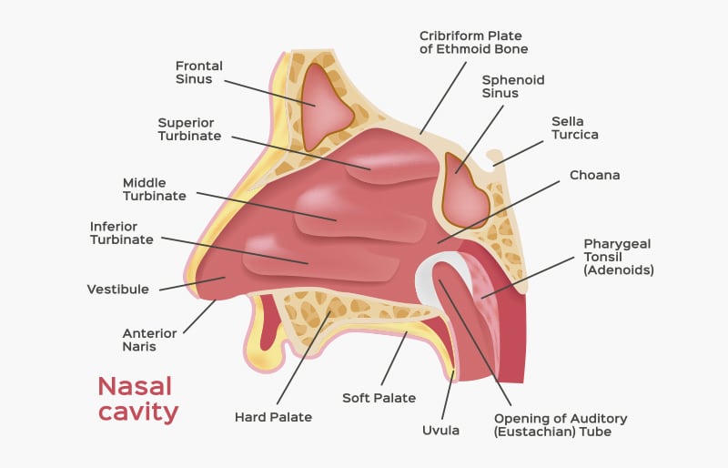
The septal cartilage is the central support structure of the nose, located between the nostrils and extending deep into the nasal cavity. It forms a key part of the nasal bridge and plays a major role in nasal shape and alignment. In rhinoplasty, surgeons often reshape or reinforce the septal cartilage to improve symmetry, support the nasal tip, and maintain proper airflow after surgery.
Upper Lateral Cartilages
The upper lateral cartilages form the middle third of the nose and connect directly to the septal cartilage. These structures help define the shape and width of the nasal bridge while supporting the internal nasal airway. During rhinoplasty, surgeons must carefully preserve this area to avoid middle vault collapse or breathing difficulties.
Nasal Bones and Facial Support Structures
These bones form the structural foundation of the nose and support the upper nasal framework during rhinoplasty and facial reconstruction procedures.
Ethmoid Bone
The ethmoid bone is a critical structure located in the upper portion of the nasal septum and forms part of the medial walls of the eye sockets. In rhinoplasty, it plays a supportive role in the overall nasal framework. This bone is often involved when harvesting septal cartilage for grafting, with surgeons preserving around 10mm of dorsal and caudal struts to maintain nasal integrity. Its strategic position and structure make it essential for both nasal function and structural reinforcement during reconstructive efforts.
Frontal Bone
The frontal bone forms the upper portion of the face and the forehead, extending down to connect with the nasal bones. It houses the frontal sinuses and contributes to the contour and projection of the upper face. In rhinoplasty, this bone provides a critical point of reference when evaluating nasal projection and facial harmony. The glabella, a key landmark located centrally between the eyebrows, is part of the frontal bone and influences the visual starting point of the nasal profile.
Frontal Process of the Maxilla
The frontal process of the maxilla is a bony projection that connects the maxilla to both the nasal bones and the frontal bone, forming part of the sidewall of the nose. It plays a key role in shaping the upper third of the nasal structure and contributes to nasal width. During rhinoplasty, this area may be involved in osteotomies—controlled bone cuts used to reshape and reposition the nasal bones. Adjusting this part of the anatomy can significantly influence the width and contour of the nasal bridge.
Maxilla
The maxilla is a foundational facial bone that forms the upper jaw, contributes to the cheek structure, and shapes part of the nasal cavity. In rhinoplasty, the maxilla plays an important role in supporting the base of the nose and influencing nasal projection. It also helps form the anterior portion of the palate and connects with the vomer and palatine bones to create the lower segment of the nasal septum. Understanding its relationship with the nasal bones and septum is key when addressing nasal balance and support.
Nasal Bone
The nasal bones form the upper third of the nose and create the structural foundation of the nasal bridge. In rhinoplasty, these bones may be repositioned through osteotomies to narrow the bridge, improve symmetry, or correct a crooked appearance while preserving overall support.
Palatine Bone
The palatine bone is a small but important structure located toward the back of the nasal cavity. It contributes to the formation of the hard palate, the floor of the nasal cavity, and part of the nasal septum. In rhinoplasty, its relevance lies in its connection to the vomer and maxilla, which together form the posterior and inferior segments of the nasal septum. Understanding this deeper anatomy is essential for structural work and for procedures that involve internal nasal support or septal angle adjustments.
Vomer Bone
The vomer bone is a thin, flat bone that forms the posterior and inferior portion of the nasal septum. It connects with the ethmoid bone above and the maxilla and palatine bones below, helping to complete the nasal septal structure. In rhinoplasty, the vomer is sometimes involved when harvesting grafting material or reinforcing the septum. Its structural role is vital in supporting the internal nasal framework and maintaining the straightness and strength of the septum.
Secure Your Safe Procedure Experience with Dr. Louis DeJoseph
Join our satisfied clients who’ve experienced safe, effective treatments.
How Nasal Anatomy Affects Rhinoplasty Surgery
Every structure in the nose works together to influence both appearance and breathing. During rhinoplasty, surgeons don’t just focus on one area; they evaluate how the nasal bones, cartilage, and septum support the entire nose as a whole.
The upper portion of the nose is shaped mainly by bone, while the middle and lower portions rely more on cartilage for structure and flexibility. Changes to the nasal bridge, tip, or septum can all affect the balance of the nose and even the airflow through the nasal passages.
For example, refining the nasal tip may involve adjusting the lower lateral cartilages, while correcting a crooked bridge may require reshaping the nasal bones. The septal cartilage is also important because it helps support the nose internally and is often used as a graft during rhinoplasty.
A successful rhinoplasty is about more than appearance alone. Surgeons must carefully preserve support and airflow while creating natural-looking results that fit the rest of the facial features.
Common Rhinoplasty Directional Terms
Rhinoplasty surgeons often use directional terms to describe the position and movement of nasal structures during surgery. Understanding these common terms can make it easier to follow discussions about nasal anatomy, tip rotation, bridge refinement, and surgical planning.
Dorsum (Dorsal)
The dorsum refers to the ridge or “back” of the nose, extending from the radix down to the nasal tip. It’s the surface most commonly reshaped during rhinoplasty to smooth out bumps, often referred to as a nasal hump, to adjust height, or refine the nasal profile. The term “dorsal” is used as an adjective to describe elements located along this ridge. Proper preservation and support of the dorsal area are crucial; otherwise, issues like saddle nose deformity may occur if too much structure is removed.
Caudal
The term “caudal” refers to a direction toward the feet or the lower part of the body. In rhinoplasty, this is often used when discussing the caudal end of the nasal septum. Modifying the caudal septum can shorten the nose or alter the rotation of the nasal tip. It plays a significant role in tip positioning, nasal length, and overall nasal projection.
Medial
“Medial” describes a position closer to the midline of the body. In rhinoplasty, it’s often used when referring to structures near the center of the nose, such as the septum.
Lateral
“Lateral” refers to a position away from the midline of the body. In rhinoplasty, it refers to structures on the outer sides of the nose, such as the alae or lateral crura.
Cephalic
“Cephalic” refers to a direction toward the head or upper part of the body. In rhinoplasty, the term is often used to describe the orientation of nasal structures, such as the cephalic positioning of the lower lateral cartilages. If these cartilages are positioned too high toward the head, it can lead to an overly rounded nasal tip or upward-curled nostrils. Cephalic trim techniques may be used to refine the tip and achieve a more balanced nasal contour.
Before and After Photos
* Each patient is unique and individual results may vary.
Work With an Expert Rhinoplasty Surgeon
Choosing the right surgeon for your rhinoplasty is essential to achieving natural-looking results that align with your facial features. A highly skilled facial plastic surgeon understands the delicate anatomy of the nose and how each adjustment impacts not only appearance but also breathing and functionality. Nose surgery, from primary rhinoplasty to revision rhinoplasty, requires precision, experience, and a keen aesthetic eye, qualities only a seasoned expert can provide.
Premier Image Cosmetic & Laser Surgery is Atlanta’s first cosmetic surgery practice center dedicated to both facial and body aesthetic procedures, including reconstructive surgery. Since 1970, we’ve set the standard for excellence by combining cutting-edge techniques with a deeply personalized approach to care. Our board-certified surgeons bring over 30 years of combined experience to every consultation and procedure, ensuring natural-looking results backed by expert skill and compassion. Call us today at 770-457-6303 or visit our contact page to schedule your consultation.